Surgery within the spinal cord is never a decision to take lightly. Intramedullary tumors arise within the cord itself, placing tumor treatment beside pathways essential to movement and sensation. These uncommon lesions warrant evaluation by a neurosurgeon with substantial experience in intramedullary surgery and an experienced neurophysiology team. Precision in a remarkably small spaceThe spinal cord is only roughly finger-width, with dimensions varying by level and individual anatomy. Microsurgical judgment must balance tumor removal with neurological function. Tumor characteristics, the dissection plane and the patient’s baseline examination shape what can safely be achieved. Many pathways. Very little room.
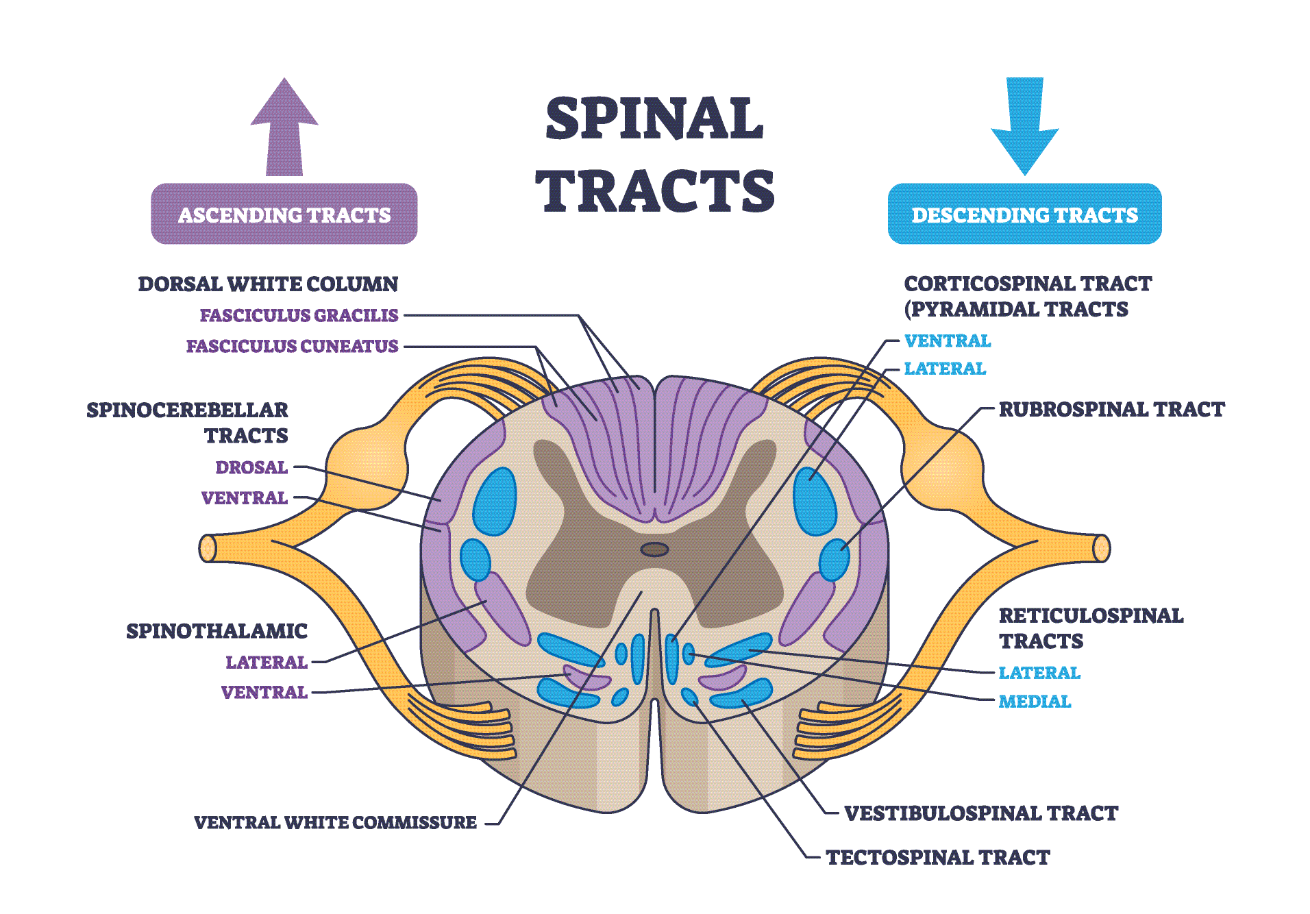
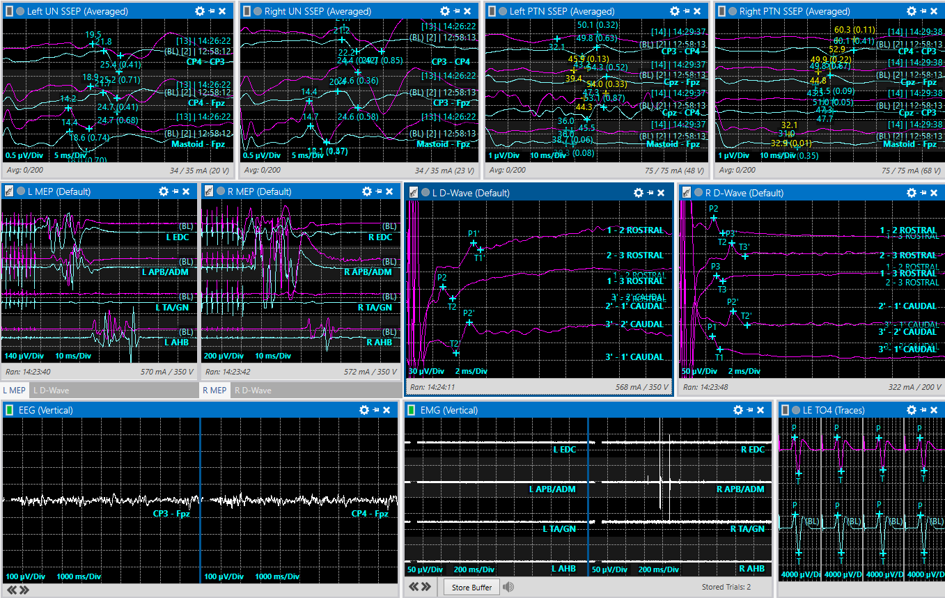
White matter surrounds the central gray matter and contains closely arranged sensory and motor tracts. Dorsal column pathways carry information about fine touch and position; anterolateral pathways carry pain and temperature signals. Descending corticospinal fibers contribute to voluntary movement. A tumor can distort this compact arrangement, making precise anatomical orientation and functional monitoring essential to surgical planning. D-wave monitoring addresses corticospinal conduction. It does not survey every pathway depicted here, which is why motor, sensory and other monitoring information must be interpreted together. Monitoring above and below the lesionD-waves assess conduction in fast corticospinal fibers after transcranial electrical stimulation. A distal (caudal) recording tracks conduction below the operative region. When feasible, a proximal (rostral) recording provides a control above it, helping the team interpret changes across the surgical region. 
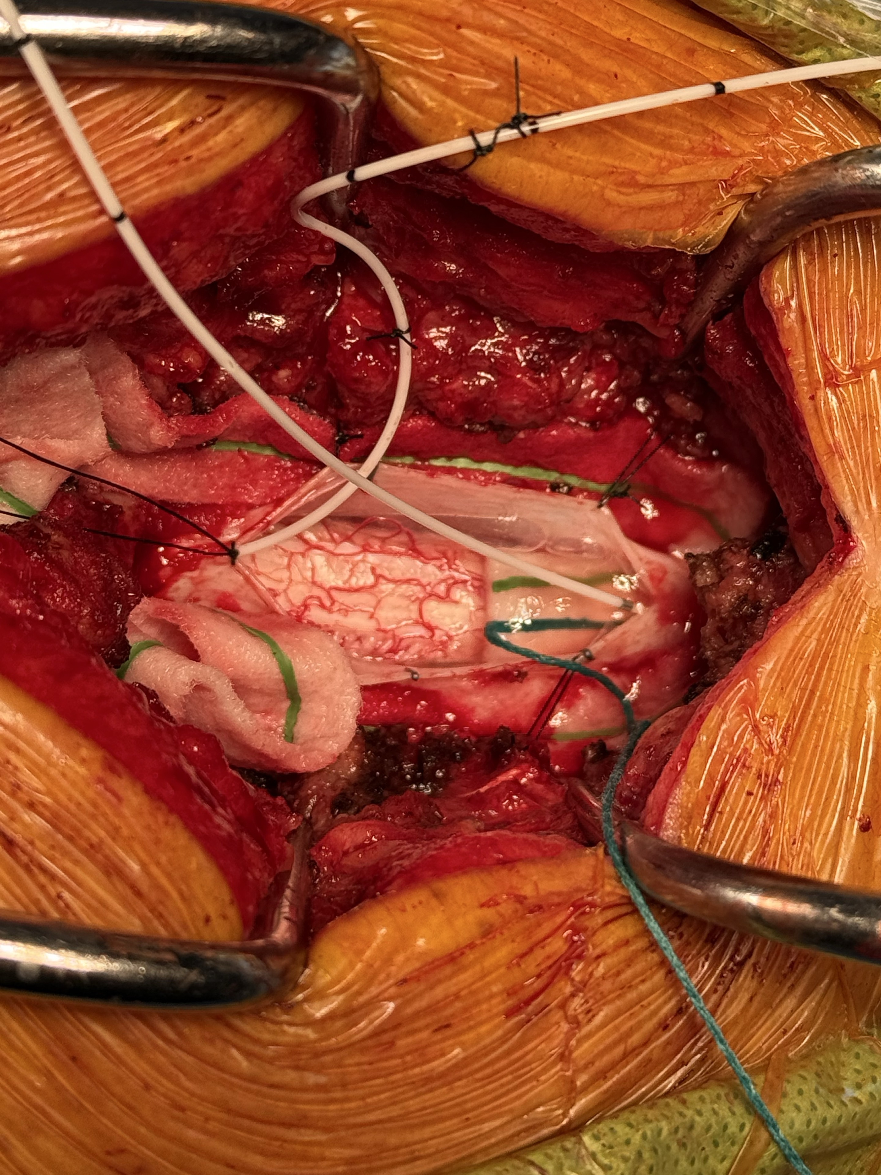
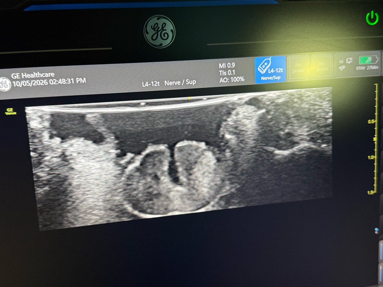
Expert interpretation is as important as the signalD-waves complement muscle motor evoked potentials and somatosensory evoked potentials; they do not replace them. Signal deterioration can prompt reassessment of surgical manipulation and physiological or technical factors. Recordability varies with lesion level, anatomy and pre-existing impairment. Preserved D-waves can be reassuring for longer-term motor function, yet temporary weakness may still occur. Monitoring supports informed intraoperative decisions; it cannot eliminate neurological risk. Inside the operating roomOperative view of the spinal cord. 

A thoughtful referralFor a suspected intramedullary tumor, seek specialist review of the imaging, neurological examination and treatment options. Ask about the surgeon’s experience with the specific lesion and the team’s approach to multimodal monitoring. |